The group working in the field of Optics and Optometry of the University of Milano-Bicocca is multidisciplinary. The main research activities concern optometry, contact lens fitting, materials science, optical systems, optics and spectroscopy for application in ophthalmic/optometric instrumentation, biology, visual perception.
Optometry
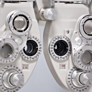
- Manual and computerized phoropters
- Automatic lensmeter
- Automatic wavefront lens analyzer
- Digital slit lamps
- Autoref / Keratometer
- Handheld autoref
- Open-field autoref
- Photorefractometer
- Digital non-mydriatic retinal camera with fundus autofluorescence (FAF)
- Non-contact Tonometer / Pachymeter
- Rebound Tonometer
- Corneal topographer – Scheimpflug Camera / Ocular aberrometer
- Open-field aberrometer
- Topographer / Biometer
- Optical coherence tomograph
- Corneal endothelial microscope
- Digital Vision Screener (VisioSmart)
- LCD vision chart
- Javal-Schiotz and Helmholtz keratometer
- Ophthalmoscopes and retinoscopes
- Osmolarity system
- Dacrioscope
- Goldmann kinetic perimeter and automated static perimeter
- Laboratory-to-classroom digital live connection
- Video centration system
- Monocular/binocular eye tracker
Optics and spectroscopy
- Spectrophotometers for UV-visible-NIR transmission and reflectivity analyses (both solutions and solid samples), both in specular configuration and by integrating sphere for the diffuse light
- UV-visible spectrometer for absorbance measurements on a few microliters of liquids
- Solar simulator light source and equipment for analyses on photochromatic lenses
- Variable-angle spectroscopic ellipsometer (200-1700 nm) for measurements of refractive index, Abbe number, and/or thickness of bulk samples, monolayer, and multilayers
- Instrumentation for photoluminescence analyses
- Instrumentation for spectrally-resolved illuminance and photometric analyses
- Spectroradiometer
- Refractometer for liquids
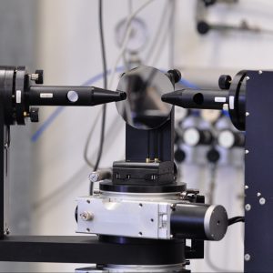
- Contrast and MTF analyses on optical systems, such as ophthalmics lenses
- Optical polarized microscope and fluorescence microscope
- Ray-tracing optical simulation techniques
Surface characterization: AFM laboratory, profilometer, tribometer
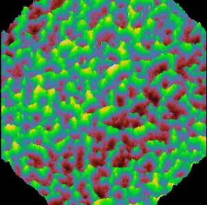
- AFM (Atomic Force Microscopy) to elaborate/model the contact lens surface morphology at micrometric and nanometric scale
- Profilometer to analyze different surfaces at macroscopic and micrometric scale
- Nanotribometer to test contact lenses in liquid
SEM interdipartimental laboratory
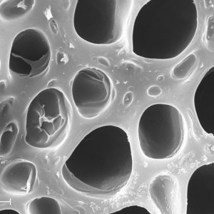
SEM (Scanning electron microscope) can produce images of different samples; for example, it can provide images of the contact lenses to study their morphology changes before and after the fitting depending on protein, lipid and maintenance solutions components absorption
NanoMat@Lab
Development of new hybrid materials for contact lenses and ophthalmic lenses and EPR (Electron Paramagnetic Resonance) analysis
and…
- DSC (Differential Scanning Calorimetry) Laboratory
- Biology Laboratory
- Organic chemistry laboratory
